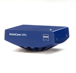
Low-intensity fluorescences, living organisms and the finest color graduations– the image quality of the AxioCam MR clearly exceeds that of other monochrome and color CCD cameras.
With a dynamic range of more than 1:2200, the highly sensitive 2/3” CCD sensor acquires up to 14 images per second in full resolution. You can then process and publish the images using the AxioVision AC imaging software provided.
For applications in fluorescence microscopy, we recommend the AxioCam MRm monochrome variant – for maximum resolution without color interpolation and without a light-reducing filter mask.
 Using our highest-performance camera you can for instance analyze processes in the fields of pathology and developmental biology. You can document your results with the best color reproduction and with every detail retained. Two switchable read-out speeds satisfy every requirement on the scale between high resolution and speed. They considerably expand the possible applications of this camera
Complete color information at every pixel
A moving sensor evaluates every detail in the red, green and blue channels. Color fringes at fine structures and edges, which occur when using cameras with a fixed sensor, are therefore avoided. Tissue discolorations can be interpreted quickly and reliably with 42 bit RGB color depth and a dynamic range of up to 1: 2500.
All details with up to 13 megapixels
With nine different resolutions you can generate images of the finest cell structures for meaningful presentation.
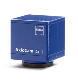
Cost-conscious scientific documentation
The models of our economically priced camera series allow you to acquire images without the time-consuming steps involved with memory cards. Thanks to the FireWire connection with speeds of up to 400 Mbit/s, the image data are transferred to your computer directly and unaltered. Whether for transmitted-light, reflected-light or fluorescence images, you will find the AxioCam IC the perfect microscope camera for your day-to-day applications. Depending on your requirements choose one of our three AxioCam IC models:
AxioCam ICc 1: color and a fast live image
The live image of this 1.4-megapixel CCD camera is refreshed up to 28 times per second. This means that you can focus through your samples smoothly and without perceptible delay. You and your employees will benefit from the color fidelity also displayed on the monitor: thus the strain of looking through the eyepiece can be avoided. Even dynamic processes or moving specimens can be monitored conveniently by several people in the live image. Thanks to its good dynamics, we recommend the AxioCam ICc 1 for images in brightfield and phase contrast in particular.
AxioCam ICc 3: higher resolution
Would you like to present or print out your images in a large format? If so, the AxioCam ICc 3 is the right camera for your laboratory. With its 3.3-megapixel color CCD it documents your samples with a resolution of up to 2080 x 1540 pixels.
AxioCam ICm 1: monochrome for bright fluorescence signals
This 1.4-megapixel monochrome CCD camera has an expanded dynamic range in relation to the color variant. As a result, you can document bright fluorescence signals with good contrast gradations and an appealing level of quality. In spite of its attractive price, the AxioCam ICm 1 has inherited some of the features of the high-end AxioCams. It allows you to digitize your image data using 12 bits/pixel and make use of functions such as black reference or hot pixel correction. The AxioCam ICm 1 is a particularly attractive option in combination with the AxioVision LE module Fluorescence Lite. Using a manual microscope, you can therefore generate fluorescence images in AxioVision more economically than ever before.
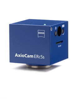
The AxioCam ERc 5s microscope camera can be operated via AxioVision in the conventional way, but it also works as a stand-alone imaging station. Neither a PC nor any software are required in order to acquire images of your samples. This ZEISS camera acquires color images with 5 megapixels and stores them on memory cards in the economical and widely used SD format. The AxioCam ERc 5s is operated simply via two easily accessible buttons: one triggers the acquisition of the image, while the other performs the automatic white balance. If you subsequently wish to transfer your images to a PC, all you need to do is insert the SD card into the memory card slot on your PC.
The AxioCam ERc 5s is compatible with all Carl Zeiss microscopes, making it possible for every microscope to be transformed, cost-effectively, into a full-fledged, mobile imaging station. This means that you can organize your laboratory processes much more flexibly than before, as you can decide which instrument you want to use to acquire images of your samples and where you do so. With our camera, troublesome tangled cables are a thing of the past.
If the requirements of your application change, or if you wish to adjust key camera parameters, simply connect the camera to your PC. Using the software supplied, you can change the color saturation, contrast, brightness and many other acquisition parameters.
With the AxioCam ERc 5s you are perfectly equipped for wide-ranging areas of application. You can perform quality controls on the move, for example. Medical and biological specimens can be documented conveniently on different microscopes. After defining the acquisition settings once on your computer, you can carry out routine tasks independently of the computer equipment at your workstation.
View smooth live images conveniently in a large format
The AxioCam ERc 5s sends your live images directly to the connected monitor or projector via a USB, AV or DVI signal. In the future you will be able to discuss your research results with your work group as you view them directly and conveniently on the monitor.
Do you have to sit at your microscope for long periods of time while you are working? If so, you are certain to benefit from the remote control supplied. Viewing your samples on the monitor means that your posture, the stand size, your working position or the viewing angle are no longer an issue. You can sit back and view your specimens in comfort.
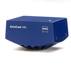
Acquire images of rapid physiological processes
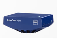
The AxioCam HS has been developed as a scientific camera for imaging applications in the fields of neurobiology, developmental biology and virology.
Analyze molecular processes and organisms in vivo
Physiological processes are acquired with a speed of up to 140 images per second. The image data is transferred immediately from the camera device to your computer without any compression losses or artifacts.
Acquire low-intensity fluorescence in several channels
With its 9.9 μm x 9.9 μm pixels, the CCD sensor of this high speed camera enables you to acquire fluorescence images with a dynamic range of 1:1800. Time lapse and Z-stack images can be generated using exposure times ranging from 1ms to 60s.
Important high end microscope peripherals such as adjustable light sources or external shutters are synchronized with the camera due to our AxioVision software. With the AxioCam HS it is possible to capture fluorescence samples with up to 8 channels and outstanding speed.
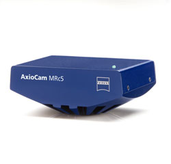
More Resolution - More Flexibility
Outstanding image quality for a wide variety of requirements in medicine and biology. With the AxioCam MRc 5, you can take advantage of 12 different acquisition modes and up to 5 megapixels resolution for your images. Thanks to the dynamic range of 1:1300 and the 36 bit RGB color depth, you can produce images with perfect color accuracy even when faced with substantial differences in brightness or reflective surfaces.
Mobile and fast
Connect the AxioCam MRc 5 with a single cable to the standard FireWire interface of your desktop or notebook. Control the complete image acquisition via this 400 megabit high speed connection and process the images instantly on your computer.